CAT Scan Contrast Agent
CAT Scan Contrast Agent — an illustrated inventions story, set in Global. 10 illustrated pages, free to read on Wonder Inventions.
Page 1
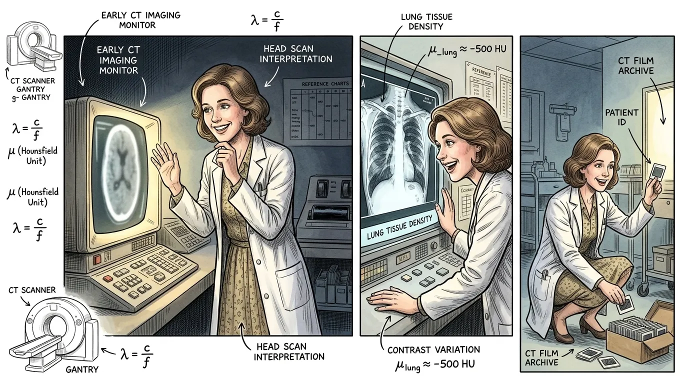
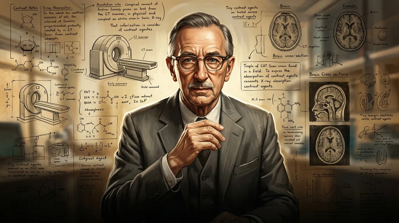
Before the advent of specialized contrast agents, medical imaging faced a formidable challenge: distinguishing between soft tissues within the human body. While X-rays revealed bones and gross anatomical structures, the subtle variations in density required for detailed soft tissue analysis remained largely invisible. Early Computed Tomography (CT) scans, pioneered by Sir Godfrey Hounsfield in the late 1960s, offered a revolutionary window into the body, but even they struggled with the nuance of internal organs, blood vessels, and pathological lesions.
""The early CT scans were remarkable," reflected Dr. Eleanor Vance, a radiologist examining a blurred scan from 1975. "We could see a tumor, yes, but its exact boundaries, its vascular supply—these critical details often remained elusive." She gestured to the faint outlines on the screen, a testament to medicine's profound limitation before enhanced visualization became possible."
Page 2
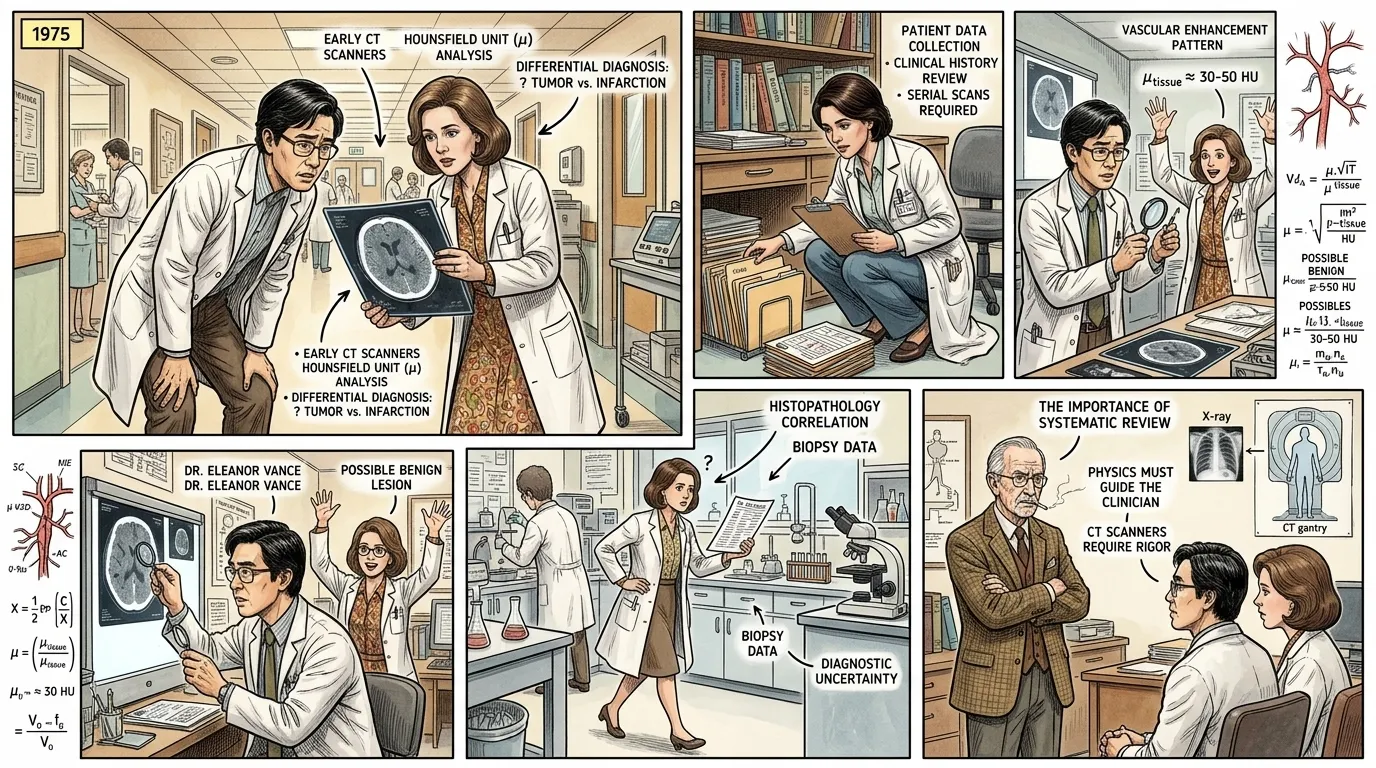
For patients, the lack of precise diagnostic tools often meant delayed diagnoses, ineffective treatments, or invasive exploratory surgeries. Tumors nestled within organs, narrowed arteries, or inflammatory processes could go undetected until they reached advanced stages. This era was marked by a clinical frustration, a knowing that diseases were lurking, yet remaining frustratingly opaque to the best medical eyes and machines of the time.
""We knew there was something wrong—the symptoms were clear," explained a concerned doctor, Dr. David Chen, speaking to a colleague in a hushed hospital hallway. "But without clearer images, pinpointing the exact location or nature of an abnormality was like searching for a shadow in the fog. How could we treat what we couldn't precisely locate?" His colleague, Dr. Anya Sharma, nodded grimly, holding a patient chart. "The human body, so complex, held its secrets too well for our current technology.""
Page 3
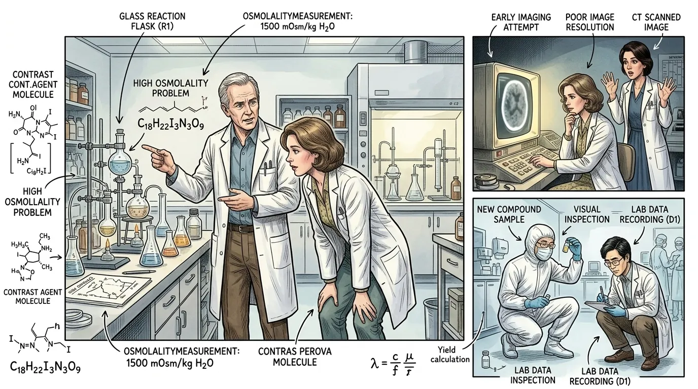
The concept of enhancing visibility for X-ray imaging wasn't entirely new; substances like barium sulfate had long been used for gastrointestinal studies. However, introducing an agent safely and effectively into the bloodstream to highlight soft tissues and blood vessels for CT scans presented a far greater biochemical challenge. Early experimental agents, primarily iodine-based, often suffered from high osmolality, meaning they had a very high concentration of particles, which could draw water from surrounding cells, leading to significant adverse reactions in patients, from nausea to severe allergic shock.
""Our initial attempts to make internal structures visible were fraught with danger," stated Dr. Marcus Thorne, a research chemist in a laboratory coat, gesturing towards beakers and flasks on a workbench. "We understood that elements with a higher atomic number, like iodine, absorbed X-rays more effectively, making them 'radiopaque.' But the toxicity! We discovered that the high osmolality of early compounds caused our patients immense discomfort and, in some cases, severe physiological stress. It was a clear demonstration of the principle that 'the road to hell is paved with good intentions,' as Samuel Johnson once famously observed, reminding us that even with the best diagnostic hopes, patient safety must remain paramount. This high osmolality was our primary barrier.""
Page 4
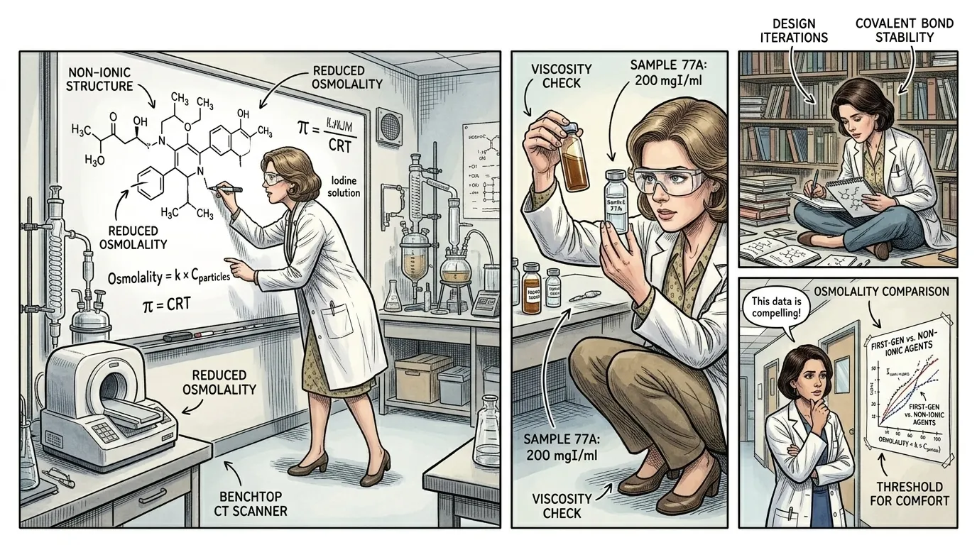
The scientific community recognized that the future of diagnostic imaging depended on developing safer contrast agents. The challenge was multifaceted: chemists needed to synthesize molecules that contained high concentrations of iodine for effective X-ray attenuation, yet remained biologically inert, meaning they wouldn't react harmfully with the body's systems. Furthermore, these compounds needed to be water-soluble, stable, and easily excretable by the kidneys. It was a complex puzzle, requiring deep understanding of organic chemistry, pharmacology, and patient physiology.
""We were essentially trying to trick the body," explained Dr. Lena Petrova, a pharmaceutical chemist, sketching chemical structures on a whiteboard. "How do you deliver a heavy atom like iodine efficiently through the circulatory system without causing havoc? The body perceives high-osmolality agents as a major imbalance. Our research focused on covalent bonding, creating molecules where the iodine atoms were tightly bound within a larger, non-dissociating structure. This was the key to reducing the free particle count and, crucially, the osmolality. We were constantly asking: how can we make this molecule invisible to the body's alarm systems, while making it highly visible to the X-rays?""
Page 5
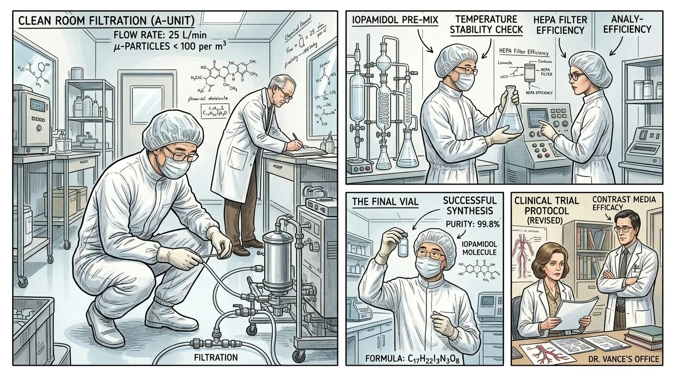
The pivotal breakthrough arrived with the development of non-ionic iodine contrast agents in the late 1970s and early 1980s. Unlike their ionic predecessors, these new compounds did not dissociate into charged particles when dissolved in blood. This significantly reduced their osmolality, making them much more isotonic with blood plasma, and dramatically lowering the incidence and severity of adverse reactions. This chemical innovation transformed the risk-benefit ratio, allowing for much wider and safer use in a broader patient population. It was a monumental step forward, unlocking the full diagnostic potential of CT imaging.
""This was it – the game-changer," stated Dr. Kenji Tanaka, a senior chemist, holding up a transparent vial of clear liquid in a clean room environment. "By creating molecules that stayed intact in solution, we bypassed the physiological shock. I remember one evening, staring at the results of our solubility tests, realizing the potential. No more dissociation, no more osmotic burden. It felt like we had finally tamed the wild beast of inorganic chemistry for the benefit of human health. This vial, containing Iopamidol, represented years of meticulous effort.""
Page 6
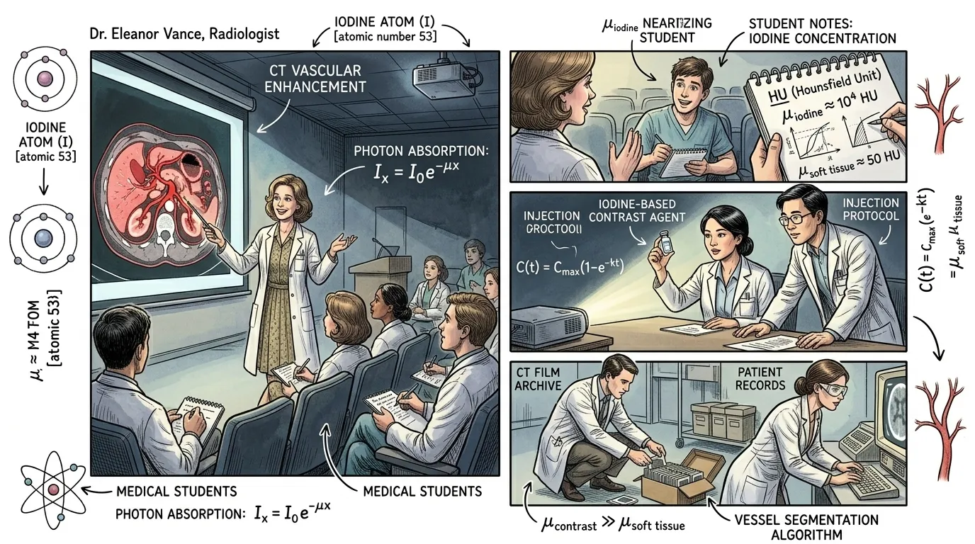
Once injected, the non-ionic iodine contrast agent travels rapidly through the bloodstream, reaching target tissues and organs. The key to its function lies in iodine's atomic properties: its relatively high atomic number means it absorbs X-rays more readily than the surrounding soft tissues, which are largely composed of lighter elements like carbon, oxygen, and hydrogen. As the CT scanner emits X-rays through the body, the areas filled with the contrast agent appear brighter on the resulting image, effectively creating a 'roadmap' of blood flow and enhancing the visibility of vascular structures, tumors, and inflammation.
""Imagine the body as a canvas, and the X-rays as light," Dr. Vance explained to a group of medical students, pointing to a detailed CT image projected on a screen. "Without contrast, everything blends. But introduce iodine, and suddenly specific areas 'light up' because the iodine atoms are avidly absorbing those X-rays. This differential absorption is what our scanner captures. The computer then translates these varying absorption levels into distinct shades of grey, revealing structures previously obscured. It's not magic; it's fundamental physics applied to biology.""
Page 7
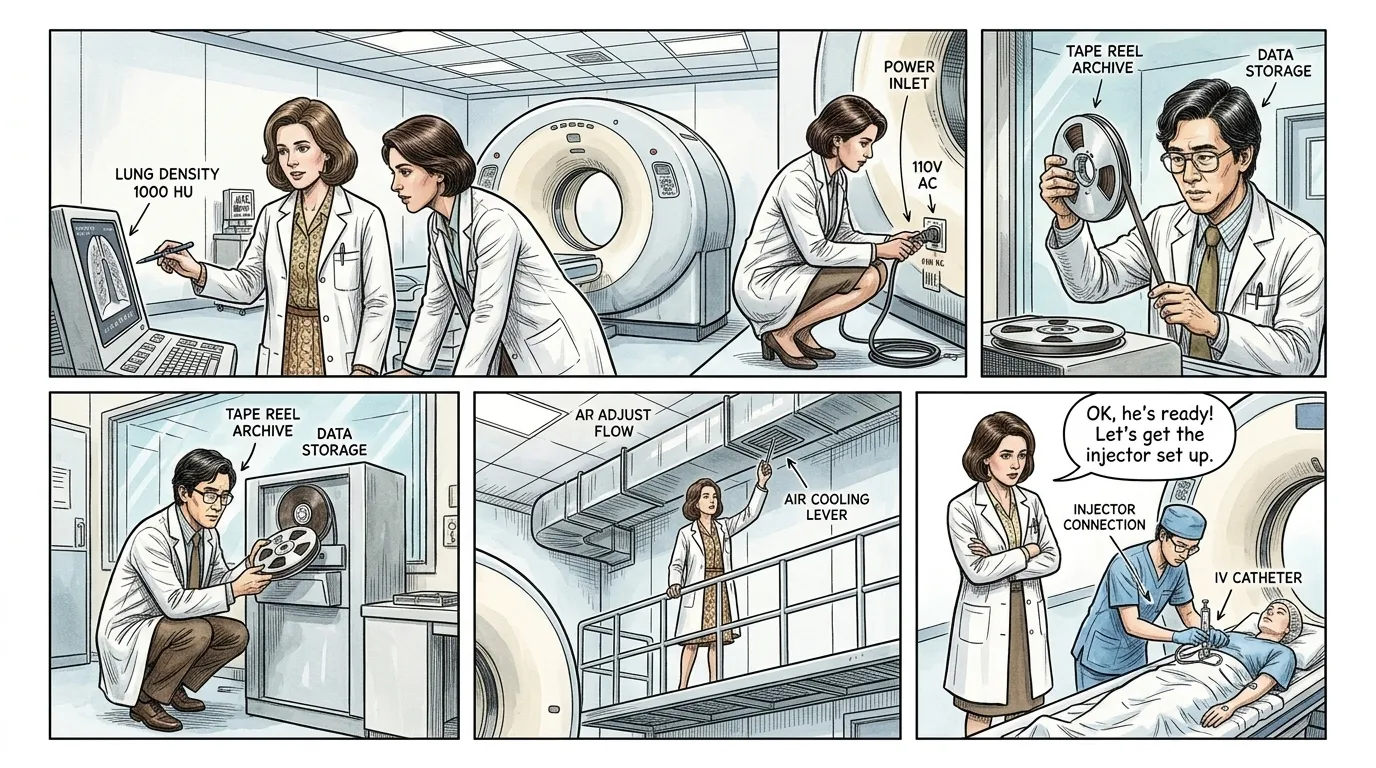
A typical contrast-enhanced CT scan begins with the intravenous injection of the agent, usually into a vein in the arm. The agent then rapidly circulates throughout the circulatory system, reaching the heart and then being pumped to all major organs. Depending on the timing of the scan, different phases of enhancement can be captured: arterial phase (highlighting arteries), venous phase (highlighting veins and organs), and delayed phase (showing accumulation in certain pathologies or excretion pathways). The kidneys then efficiently filter the contrast agent from the blood, and it is excreted from the body, typically within 24 hours.
""The timing of our scan is critical," clarified Dr. Chen, observing a technician preparing a patient for a CT scan. "Once the agent is injected, we monitor its progression. If we want to evaluate arterial blockages, we scan during the arterial phase, just moments after injection. For liver lesions, a venous phase might be more informative. It's like a meticulously choreographed dance within the body, each step revealing a different aspect. Our precision ensures we capture the information at its most diagnostic peak." The technician nodded, preparing the automated injector."
Page 8
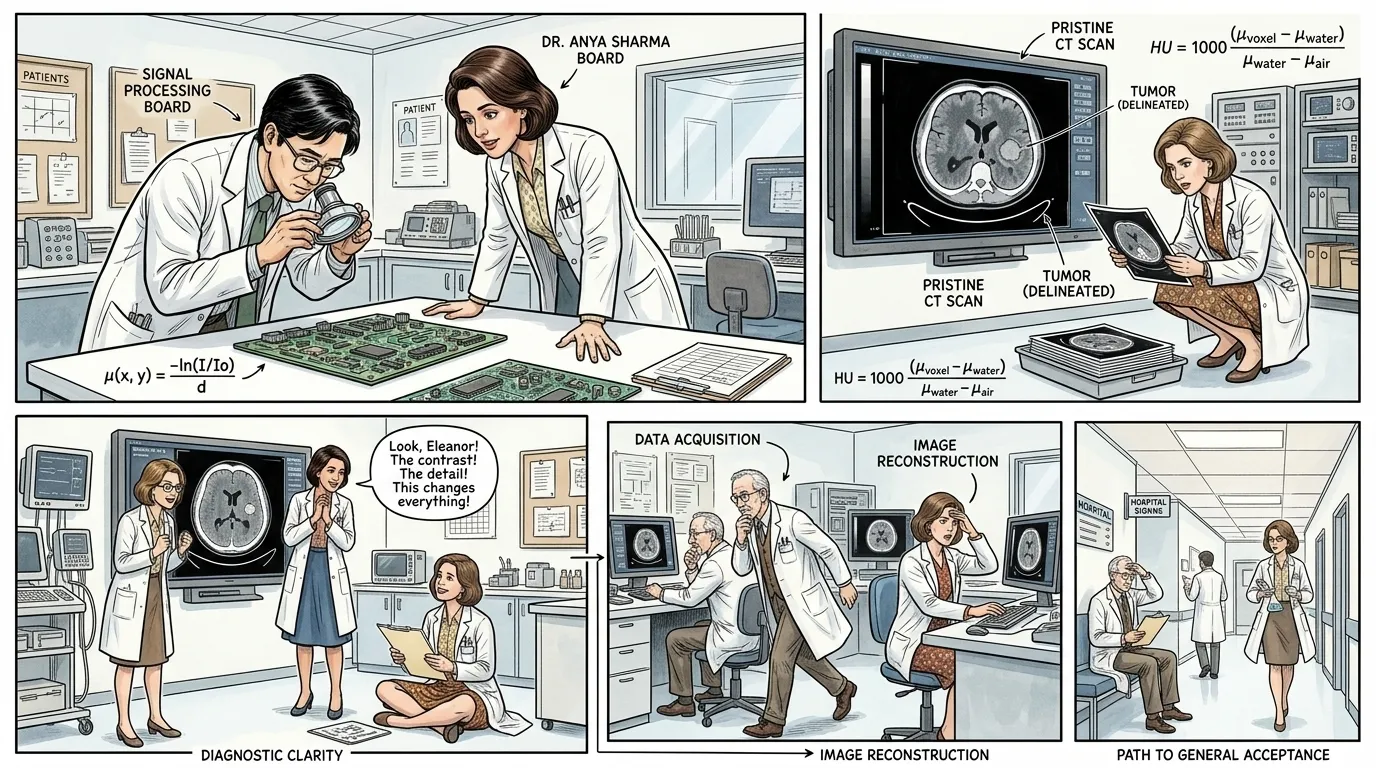
The introduction of safe, non-ionic contrast agents revolutionized diagnostic medicine. Doctors could now visualize tumors with unprecedented clarity, assess the extent of traumatic injuries, detect internal bleeding, and accurately map vascular diseases. This capability dramatically improved surgical planning, allowed for earlier detection of cancers, and enabled more precise monitoring of treatment effectiveness. The earlier issues of high osmolality and patient side effects, which had hindered widespread adoption, were largely overcome, making contrast-enhanced CT a routine and indispensable tool.
""With these new agents, we gained true clarity," exclaimed Dr. Anya Sharma, reviewing a pristine, high-contrast CT scan on a monitor. "No more guesswork, no more 'shadows.' We can now precisely locate and characterize pathology. 'The greatest danger for most of us is not that our aim is too high and we miss it, but that it is too low and we reach it,' as Michelangelo famously said. We aimed for safety and clarity, and achieved it, overcoming the osmolality issues that plagued our predecessors. This technology empowers us to provide vastly superior patient care." She pointed to a clearly delineated tumor on the screen."
Page 9
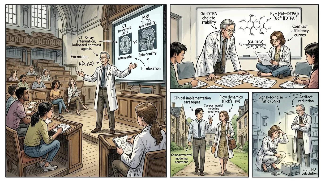
The success of iodine-based contrast agents for CT scans inspired parallel developments in other imaging modalities. For Magnetic Resonance Imaging (MRI), gadolinium-based contrast agents were developed, operating on a different principle by altering the magnetic relaxation times of protons in tissues. This expansion meant that the fundamental concept of 'contrast enhancement' transcended specific imaging technologies, providing doctors with an even broader array of tools to visualize and diagnose. From oncology to cardiology and neurology, contrast agents became integral to modern medicine, continually refined for greater safety and specificity.
""The principle of contrast enhancement didn't stop with CT," Dr. Marcus Thorne explained to a new generation of researchers in a university lecture hall. "It evolved. For MRI, we needed something different—paramagnetic ions like gadolinium. These agents work by subtly influencing the water molecules around them, altering the magnetic signals. It was a new frontier, a different chemical challenge, but the goal remained the same: to reveal the invisible. This constant drive for improvement is the essence of scientific progress." He gestured to a slide showing a comparison of CT and MRI contrast principles."
Page 10
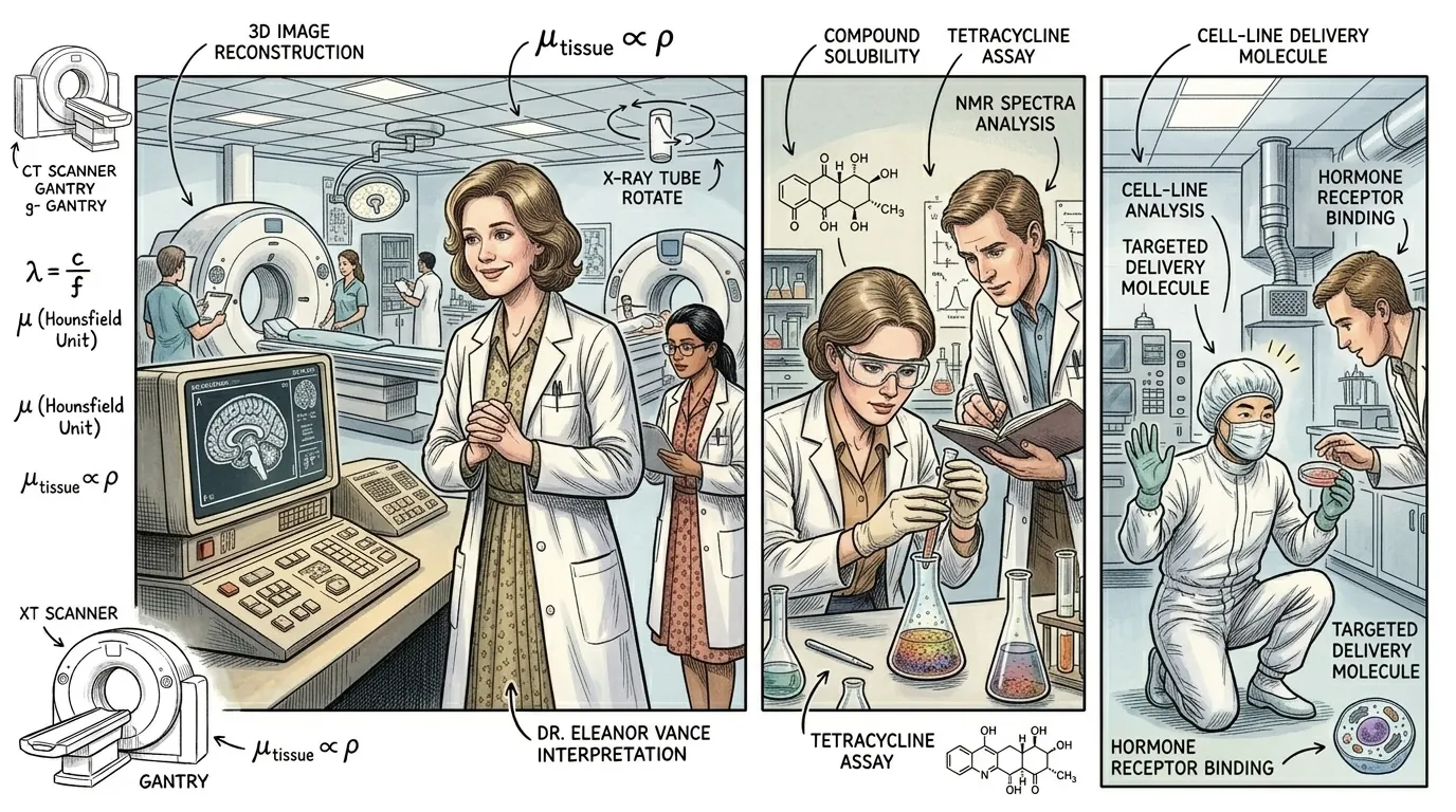
Today, CAT Scan Contrast Agents, along with their MRI counterparts, are indispensable tools in modern medicine, saving countless lives by enabling early and accurate diagnoses. They have transformed the management of cancer, cardiovascular disease, stroke, and inflammatory conditions. Ongoing research continues to develop even safer agents with enhanced targeting capabilities, leading to personalized medicine and more effective therapies. The journey from crude, toxic compounds to highly refined, non-ionic formulations stands as a testament to persistent scientific inquiry and its profound impact on human health, perpetually pushing the boundaries of what is visible within the human body.
""The impact is immeasurable," concluded Dr. Eleanor Vance, standing in a bustling, state-of-the-art radiology department. "Every day, these agents help us identify a tumor early, confirm a stroke, or guide a life-saving surgery. They allow us to see with a precision that was once unimaginable. It's not just about images; it's about informed decisions, tailored treatments, and ultimately, giving patients the best chance at life.""
About this story
- Location: Global
- Audience: general readers
Questions and answers
Read Wonder Inventions on your phone
Wonder Inventions is available on Android. Get Wonder Inventions on Google Play.