Biopsy Needle
Biopsy Needle — an illustrated inventions story, set in Global. 10 illustrated pages, free to read on Wonder Inventions.
Page 1
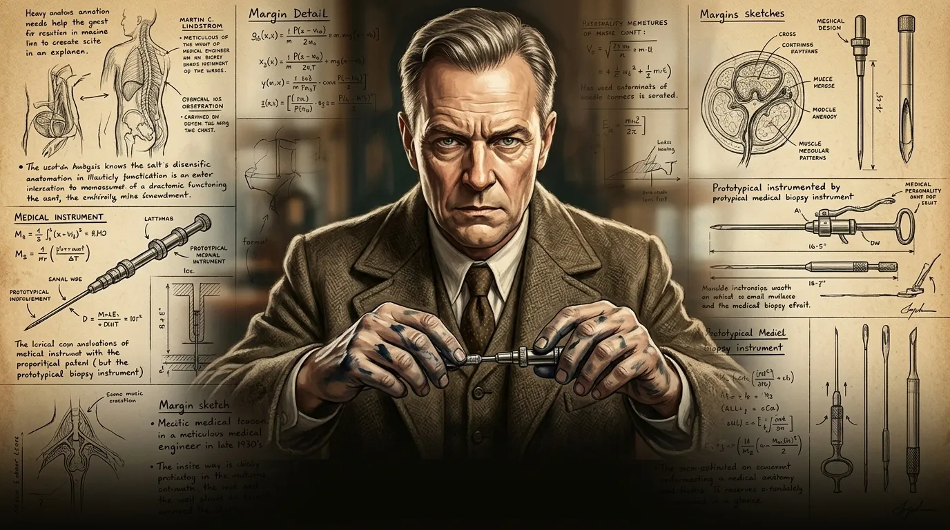
Before the mid-20th century, the landscape of internal disease diagnosis was fraught with uncertainty and invasive procedures, leaving doctors and patients alike in a perilous state of doubt.
""We face a formidable adversary in diseases hidden deep within the body," observed Professor Martin C. Lindstrom, his gaze fixed on intricate anatomical charts. "As the renowned scientist Marie Curie once remarked, 'Nothing in life is to be feared, it is only to be understood.' For us, in medicine, that understanding begins with seeing the unseen, precisely and without doubt, and our current methods are simply inadequate to achieve this with the necessary accuracy.""
Page 2
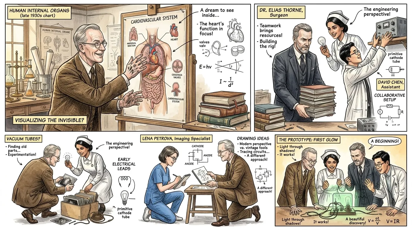
For centuries, confirming diseases like cancer within organs often necessitated extensive, high-risk exploratory surgery, a procedure that itself could imperil a patient's life or delay critical treatment.
""Consider the dilemma," began Dr. Anya Sharma, a sharp-featured woman in her early 30s with dark, pulled-back hair, wearing a starched nurse's uniform, pointing to a diagram of a human torso. "To merely identify a tumor, we had to cut deeply, sometimes through layers of healthy tissue, and even then, the small tissue fragments we managed to retrieve were frequently crushed and distorted." Dr. Elias Thorne, a serious surgeon in his 50s with a neatly trimmed grey beard and stern brown eyes, nodding grimly, added, "The fragmentation of samples rendered many diagnoses inconclusive, forcing us back to the operating table, or worse, delaying life-saving interventions.""
Page 3
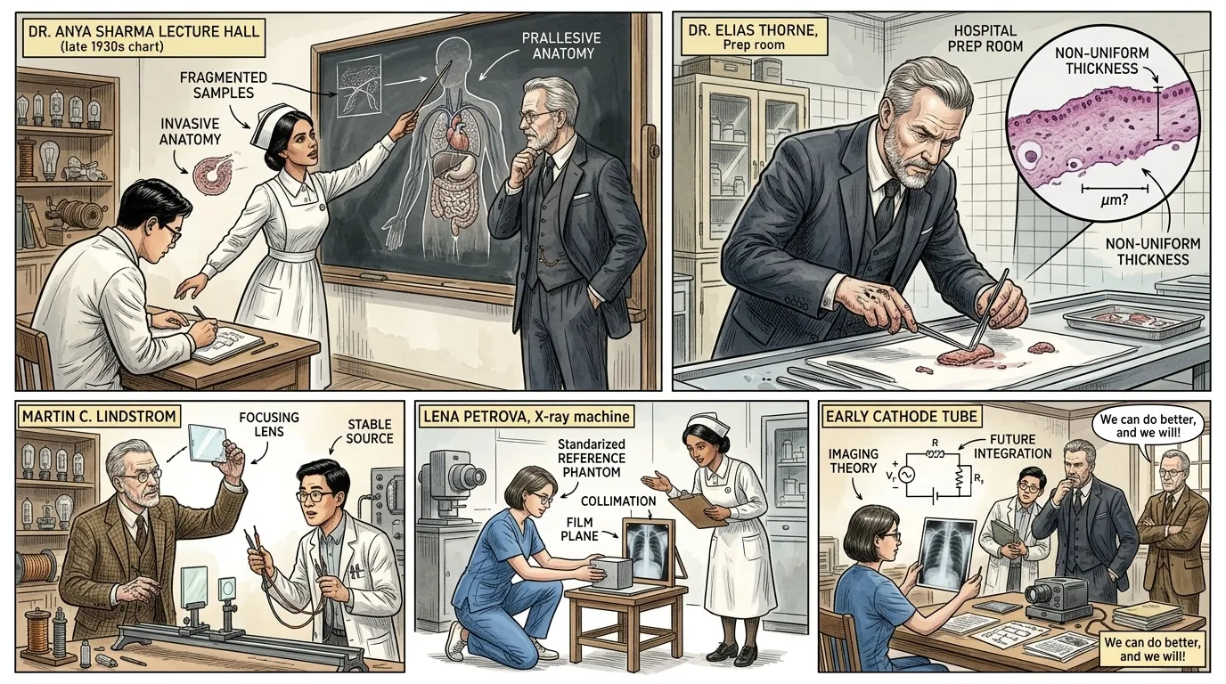
Previous attempts to gather tissue non-surgically involved simple hollow needles, similar to those used for injections, but these often struggled to extract substantial, undamaged samples from solid organs.
""The primary challenge wasn't just penetration, but precise extraction," explained Dr. Sharma, holding up a standard hypodermic needle. "While these could reach the target, their basic design either sheared off insufficient material or, more often, compressed the delicate cellular structure, rendering it useless for microscopic analysis." Dr. Thorne gestured to a large chalkboard filled with anatomical sketches and notes. "We needed a mechanism that could not only pierce but also cleanly cut a cylindrical core of tissue, preserving its architecture, a task far beyond the capabilities of simple aspiration.""
Page 4
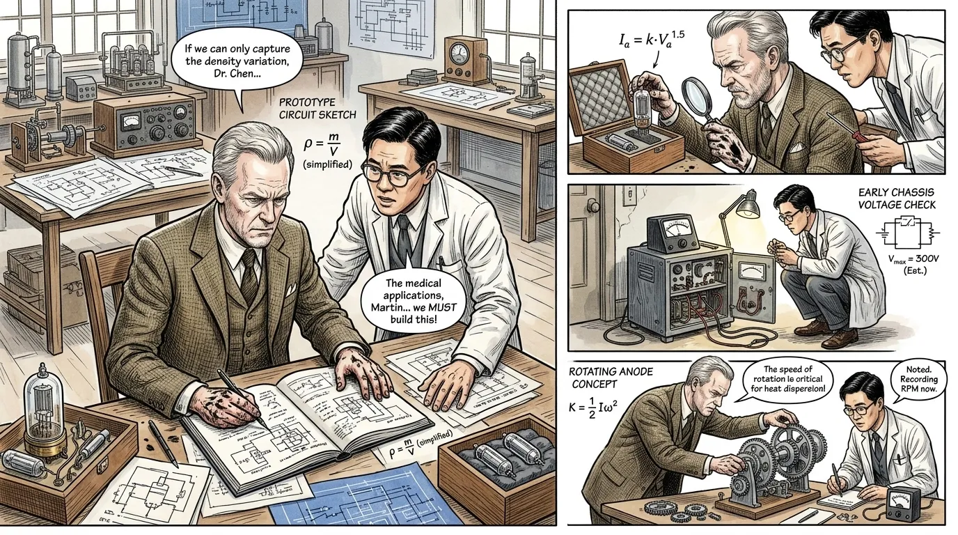
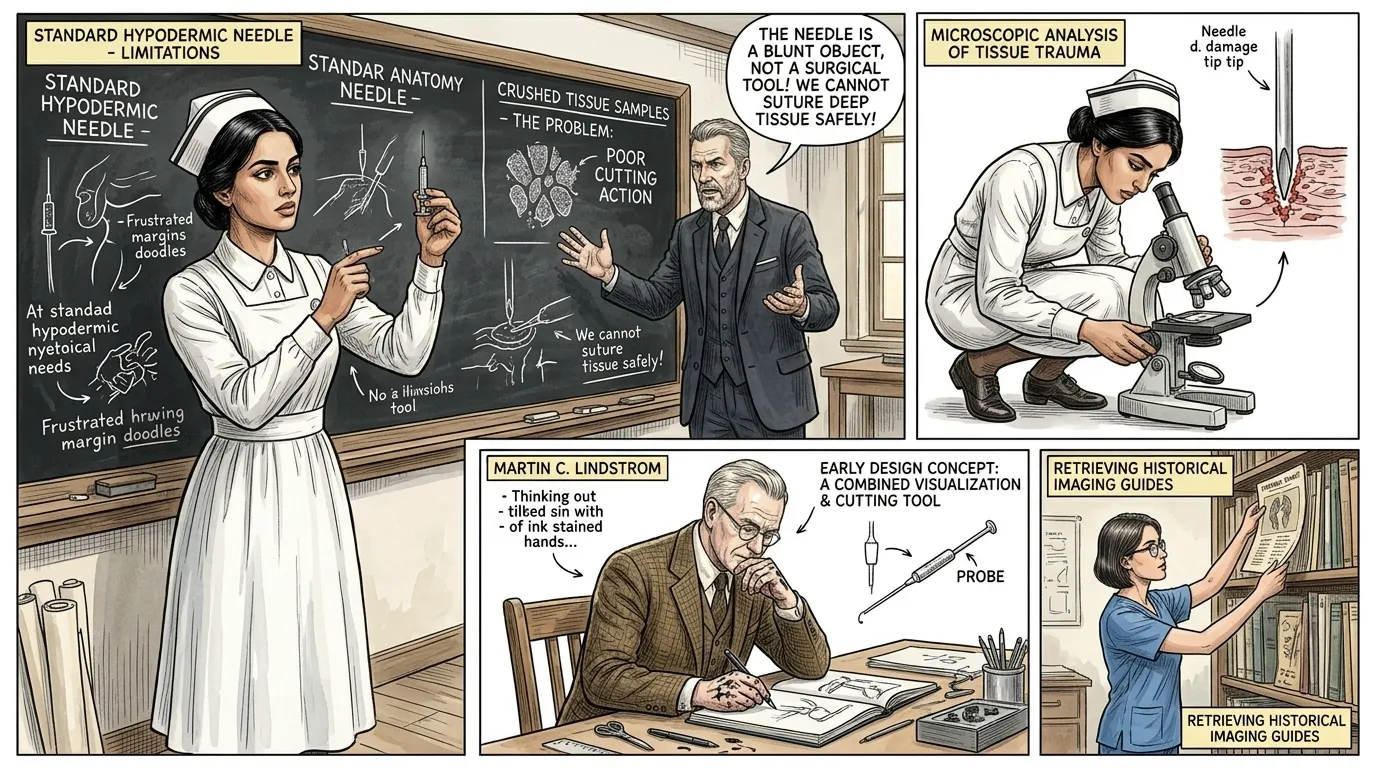
Driven by the urgent need for better diagnostic tools, Martin Lindstrom, a medical engineer and inventor, began to meticulously study tissue mechanics and instrument design in the late 1930s.
""The human body demands a delicate touch, yet decisive action," Lindstrom mused aloud, sketching furiously in a notebook filled with technical drawings. "Every crude instrument risks more harm than good, and imprecise diagnosis can be a death sentence; the sheer human cost of misdiagnosis fueled my resolve to find a better way." A junior colleague, Dr. David Chen, a young man in his 20s with short, dark hair and glasses, wearing a lab coat, nodded, stating, "We must create a tool that respects the tissue, allowing us to see its truth without destroying it.""
Page 5
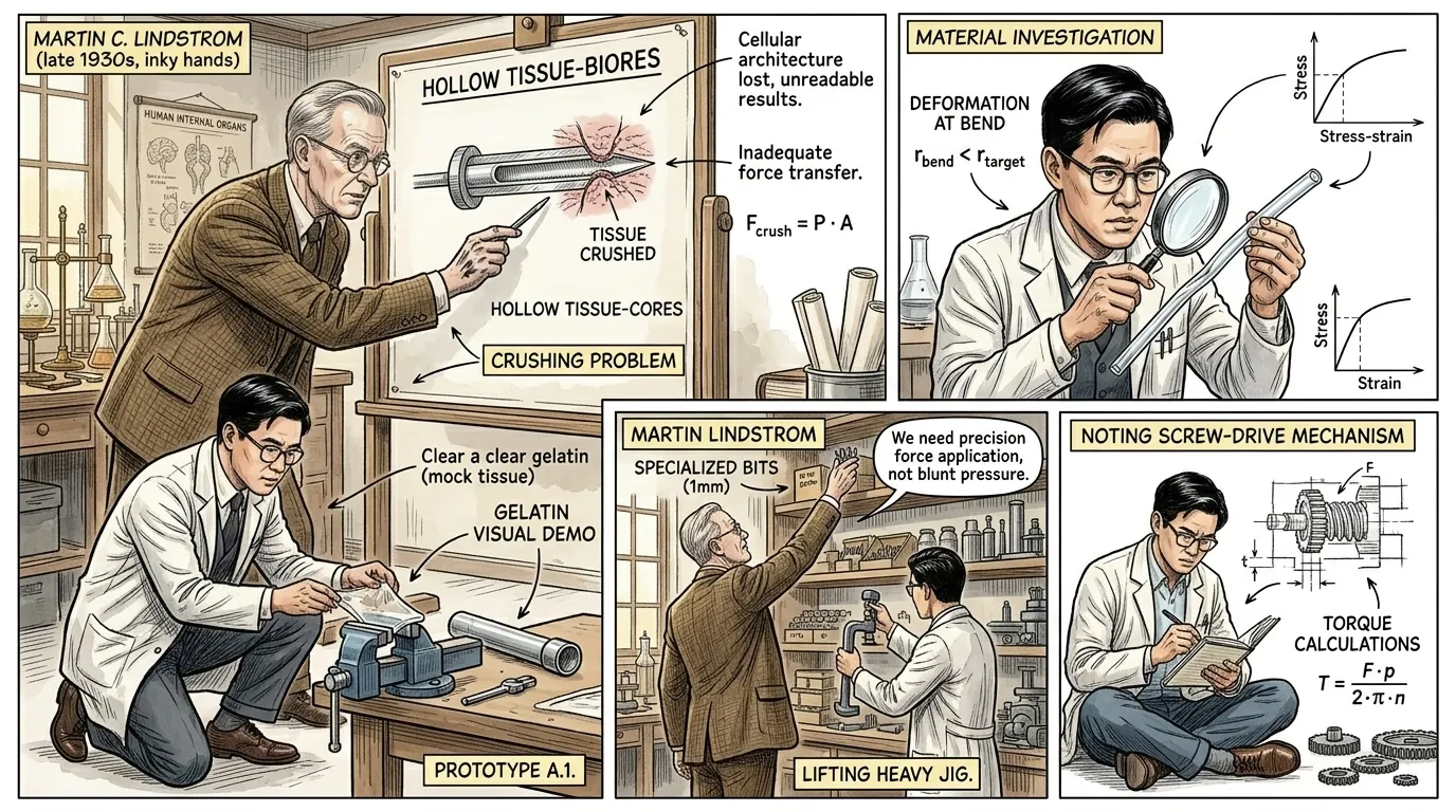
Lindstrom's initial prototypes, which mostly consisted of simple hollow tubes, consistently failed to yield intact tissue cores, either collecting insufficient material or damaging the delicate cellular structures during extraction.
""The tissue simply folds or crushes against the blunt interior of a hollow tube," Lindstrom explained, pointing to a cross-section diagram of an attempted biopsy. "We need a way to cleanly isolate a target section before extraction, not just scoop it." Dr. Chen chimed in, "Imagine trying to cut a perfect cylinder from gelatin with an open pipe – it squashes; we need a blade that defines the core before it's withdrawn, preventing distortion.""
Page 6
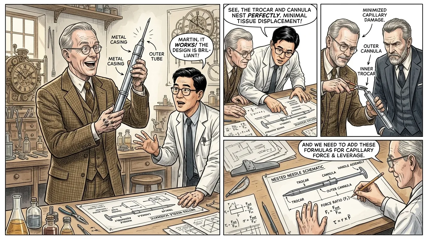
The pivotal breakthrough came with the conception of the Vim-Silverman needle in 1939 by Martin C. Lindstrom, later refined by Arthur J. Vim and Daniel Silverman, which introduced a revolutionary nested design.
""The solution lay in dual action," Lindstrom announced, holding up a prototype of his new design, a gleaming metal instrument. "We employ an inner, split stylet designed to spear and hold the tissue, and an outer cannula that slides over it, precisely cutting the core." Dr. Chen, examining a detailed blueprint, exclaimed, "It's ingenious! The split stylet anchors the tissue, and the sliding cannula acts like a miniature surgical knife, ensuring an undisturbed sample.""
Page 7
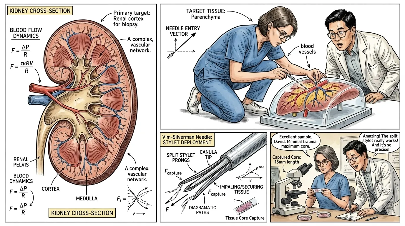
The operation of the Vim-Silverman needle is a marvel of mechanical precision, beginning with the careful insertion and advancement of its inner component.
""First, the needle assembly is inserted through the skin and guided toward the target tissue," explained a medical instructor, Dr. Lena Petrova, a focused woman in her late 40s with short, grey-streaked brown hair and practical glasses, wearing blue medical scrubs, demonstrating with a training model. "Once positioned, the inner split stylet is advanced beyond the outer cannula; its two sharp prongs splay slightly, impaling the tissue we wish to sample, meticulously securing it in place." Dr. Chen observed, "This initial capture is crucial; it prevents the tissue from simply pushing away as the cutting element approaches.""
Page 8
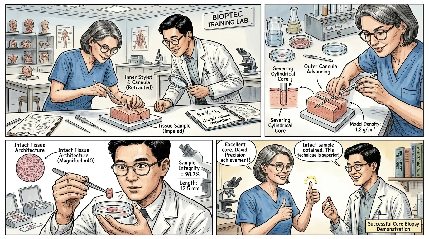
Following the impalement, the outer cannula is smoothly advanced, precisely severing a cylindrical core of tissue around the captured portion, a critical step that ensures an intact, diagnostic-quality specimen.
""This is where the genius of the design truly shines," Dr. Petrova articulated, completing her demonstration. "With the tissue firmly impaled by the stylet, the outer cannula is then slid forward over the stylet, effectively acting as a miniature cylindrical blade. It cleanly excises a core of tissue, which is then safely contained within the needle assembly, providing an undisturbed sample." Dr. Chen, looking at the excised core from the model, remarked, "This mechanism directly overcomes the fragmentation we observed with earlier methods, giving us the intact architecture vital for accurate pathological examination.""
Page 9
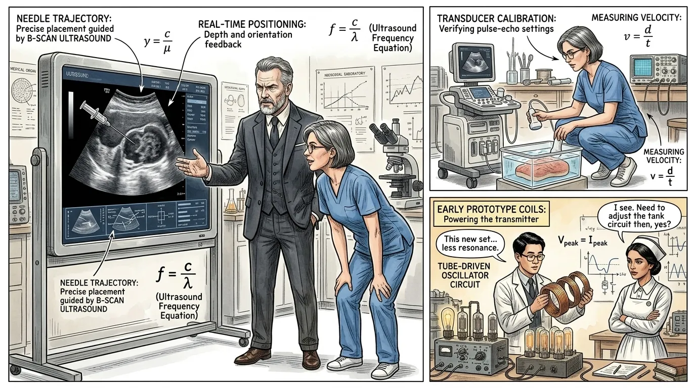
The Vim-Silverman needle quickly became a cornerstone of diagnostic medicine, but its true potential expanded exponentially with the advent of imaging technologies that allowed for precise real-time guidance.
""The initial biopsy needle was revolutionary, but blind insertions still carried risks," Dr. Thorne explained, standing before a large screen displaying an ultrasound image. "The integration of ultrasound and CT guidance transformed biopsy from a somewhat imprecise art into a highly accurate, targeted procedure, minimizing complications." Dr. Petrova added, "Now, we can visualize the needle's path in real-time, confirming its position within millimeters of the target, and expanding diagnostic capabilities to previously inaccessible areas.""
Page 10
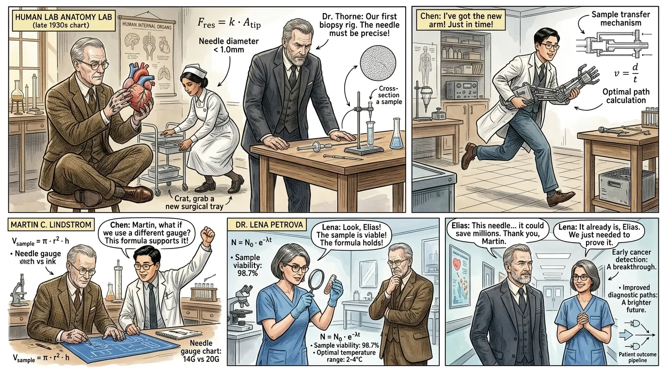
From its humble beginnings as a solution to a critical diagnostic problem, the biopsy needle has evolved into an indispensable tool, fundamentally reshaping the landscape of medical care.
""The biopsy needle has saved countless lives," Dr. Thorne stated, reflecting on its enduring impact. "It has ushered in an era of early and precise diagnosis, allowing targeted treatments to begin sooner, and often less invasively." Dr. Petrova concluded, "This elegant instrument empowers medical professionals worldwide to confront disease with unparalleled clarity, turning uncertainty into understanding, and fear into hope for millions.""
About this story
- Location: Global
- Audience: general readers
Questions and answers
Read Wonder Inventions on your phone
Wonder Inventions is available on Android. Get Wonder Inventions on Google Play.