CT Scanner
CT Scanner — an illustrated inventions story, set in Global. 10 illustrated pages, free to read on Wonder Inventions.
Page 1
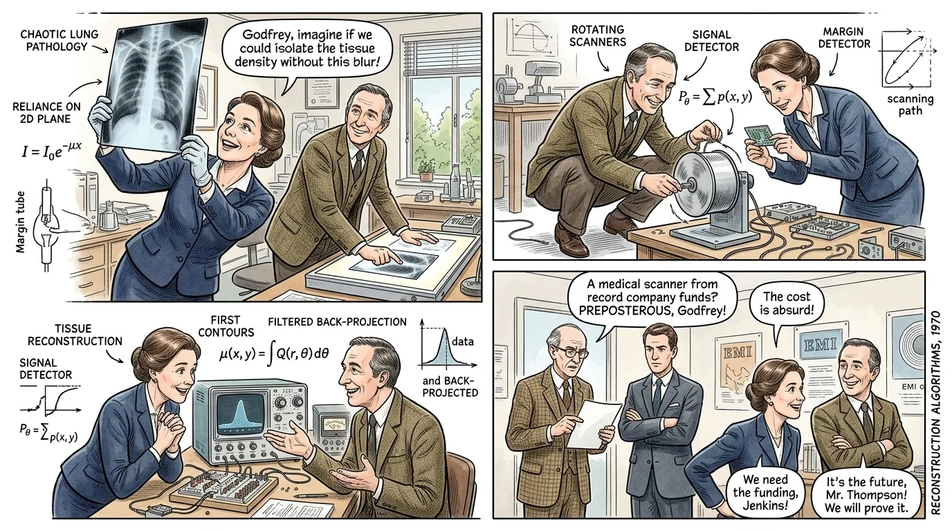
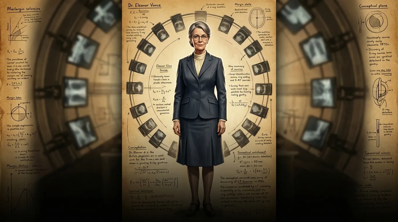
Before the Computed Tomography (CT) Scanner, diagnosing internal conditions was a fraught endeavor. Traditional X-rays provided only two-dimensional shadowgrams, a chaotic overlay of bones, organs, and soft tissues. Deep-seated tumors, subtle bleeds, or complex fractures could easily remain hidden, leaving doctors to make critical decisions with incomplete information.
"A distinguished British physician in his late 50s, Dr. Eleanor Vance, shakes her head, holding up a dense X-ray film. "It's like looking through a dense fog, isn't it? As Leonardo da Vinci once said, 'The human foot is a masterpiece of engineering and a work of art.' How true, yet how frustratingly opaque when we try to peer within it without proper tools. This overlapping tissue makes it impossible to discern the true extent of the lesion!" Across the room, Dr. Godfrey Hounsfield observed her frustration, a silent resolve forming in his mind regarding the limitations of current diagnostic techniques."
Page 2
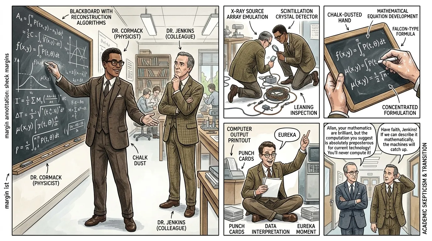
The problem of seeing through superimposed structures wasn't new. Decades earlier, in the late 1950s, South African-American physicist Allan Cormack, working at Tufts University, pondered this very challenge. He realized that if one could gather enough X-ray projections of an object from different angles, mathematics could be used to reconstruct a precise cross-sectional image. This was the fundamental theoretical 'knowledge' that would later underpin the CT scanner.
"Dr. Allan Cormack, a studious South African-American physicist in his early 50s, with thick dark-rimmed glasses and neatly combed dark brown hair, enthusiastically gestures at a complex set of equations chalked onto a large blackboard. He explains to a skeptical academic colleague, Dr. Jenkins, who listens with folded arms. "The beauty of it, Professor, is that a body's internal structure can be inferred not by staring at its surface, but by analyzing how radiation passes through it from every conceivable angle. As Pythagoras taught us, 'Numbers have a way of taking a man by the hand and leading him down the path of reason.' The raw data, after all, contains the blueprint!" Dr. Jenkins nods slowly, still pondering the sheer computational magnitude implied."
Page 3
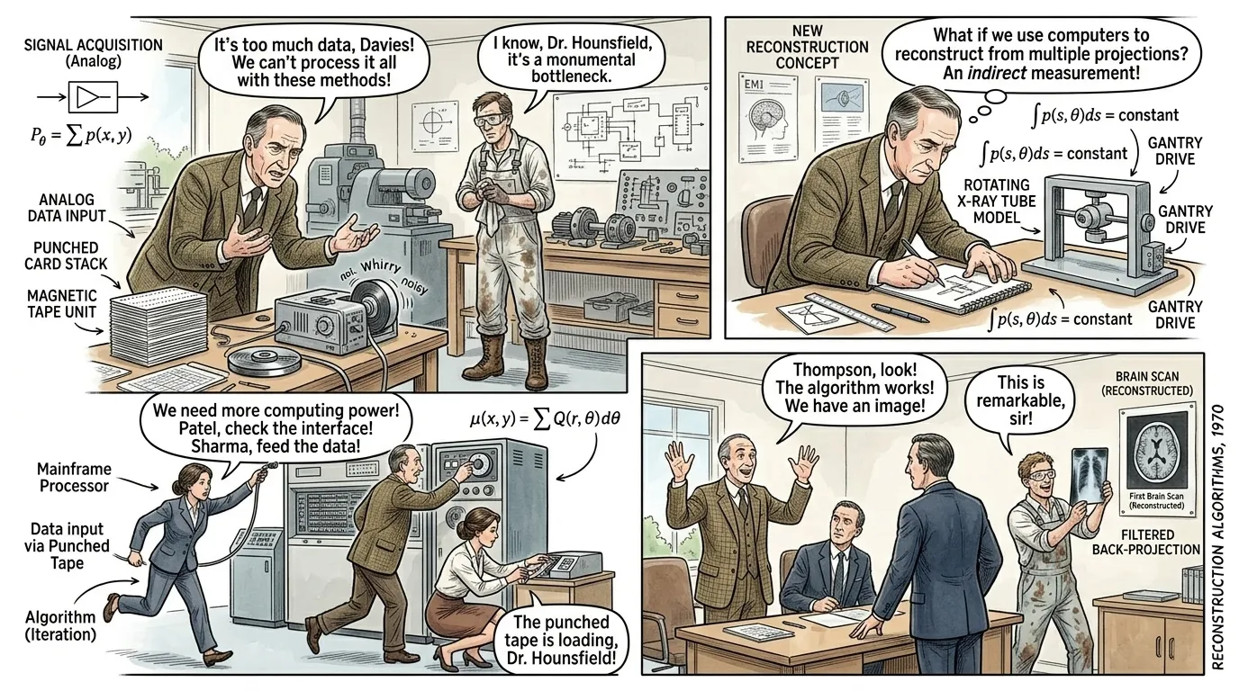
Cormack's mathematical insights provided the theoretical framework, but translating it into a working device was a monumental task. The sheer volume of X-ray data needed, and the complex calculations required to reconstruct images, far exceeded the capabilities of early analog computers. Godfrey Hounsfield, independently approaching the problem at EMI, faced these immense practical hurdles.
"Dr. Godfrey Hounsfield, now in his early 50s, with a more pronounced receding hairline but the same focused gaze, leans over a bulky, rudimentary prototype in a cluttered EMI workshop, muttering to his technician, Mr. Davies. "The analog approach simply won't suffice, Mr. Davies. Each projection is a line of data, and combining them all with mechanical filters is proving... intractable. As Thomas Edison famously said, 'I have not failed. I've just found 10,000 ways that won't work.' We've found far too many 'won't works' with this brute-force method!" He gestures at a stack of punched cards and a noisy, whirring tape drive. Mr. Davies, a practical engineer in his 40s, shakes his head in agreement, wiping grease from his hands."
Page 4
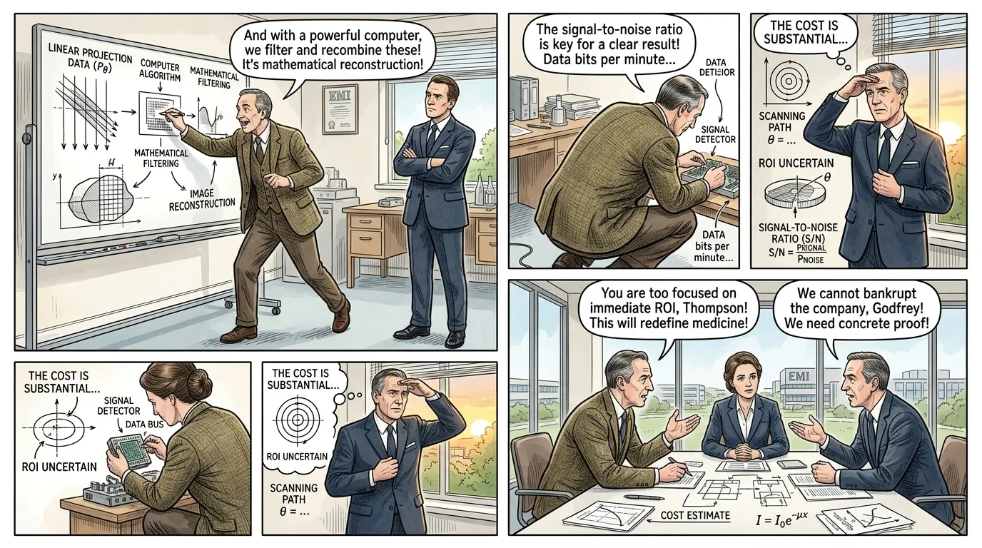
The true breakthrough for Hounsfield came with a pivotal shift in thinking: abandoning analog methods for digital computing. As digital computers became more powerful and accessible in the late 1960s, he realized their potential to process the vast amounts of X-ray attenuation data needed for image reconstruction, transforming Cormack's abstract math into a tangible reality.
"In a sparsely furnished EMI office, Dr. Godfrey Hounsfield paces excitedly, explaining his new vision to a skeptical EMI executive, Mr. Thompson. "Imagine, Mr. Thompson, we don't need a physical filter! We can mathematically filter and reconstruct the image using a digital computer. We feed the X-ray readings, hundreds of thousands of them, into a machine that performs Cormack's equations at incredible speed!" He points emphatically at a diagram he's sketched on a whiteboard illustrating digital data processing. "As Arthur C. Clarke famously posited, 'Any sufficiently advanced technology is indistinguishable from magic.' This, sir, feels like magic compared to our old methods, but it's pure mathematics and digital logic!" Mr. Thompson raises an eyebrow, considering the investment."
Page 5
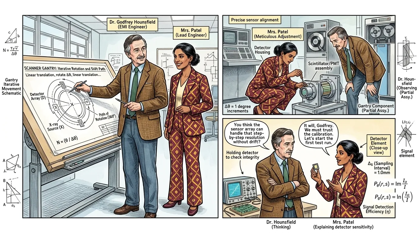
With EMI's support, Hounsfield embarked on building the first working prototype. His design featured a single X-ray source and a single detector positioned diametrically opposite each other, which would traverse linearly across the patient, taking readings, then rotate slightly, and repeat the scan. This iterative process, though slow, was revolutionary.
"Dr. Hounsfield, now overseeing a team of engineers, points to a schematic of the gantry's movement. "The X-ray tube and detector move in tandem, scanning across the patient, taking hundreds of attenuation measurements. Then, the entire assembly rotates by just one degree, and we scan again. We repeat this 180 times for a full sweep!" He turns to his lead engineer, Mrs. Patel, a sharp woman in her 30s with her hair tied back, who is adjusting a sensor. "It's painstaking, Mrs. Patel, but each tiny data point is vital. As Voltaire once observed, 'No problem can withstand the assault of sustained thinking.' We are proving that, one precise rotation at a time!" Mrs. Patel smiles, tightening a screw on the detector housing."
Page 6
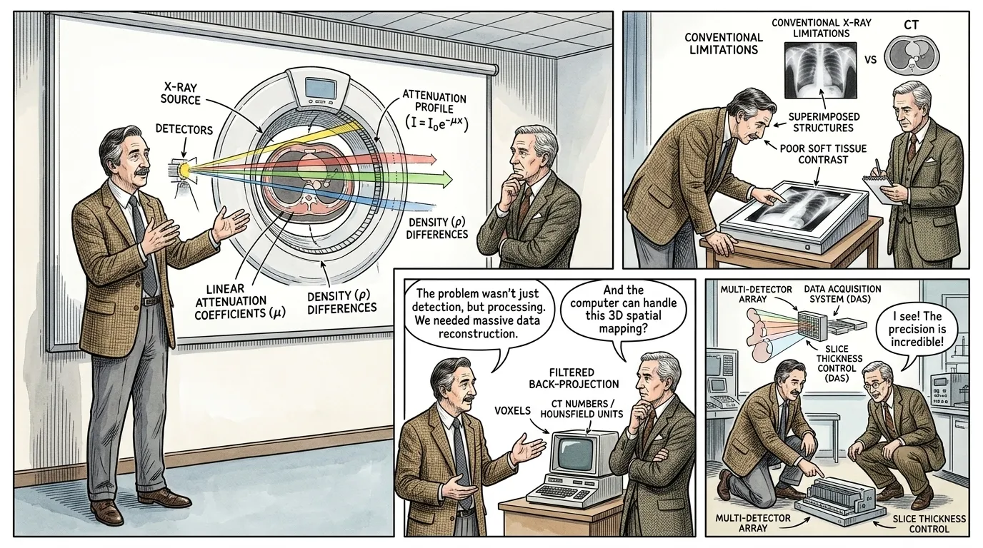
The CT scanner's operation relies on a fundamental principle: different tissues absorb X-rays to varying degrees. Bone absorbs more than soft tissue, which absorbs more than air. The scanner measures these tiny variations in X-ray attenuation as the beam passes through a specific cross-section of the body.
"Dr. Hounsfield, standing next to a sophisticated diagram of the CT gantry, explains the process to a visiting medical professor, Dr. Miller. "Think of it this way, Dr. Miller: an X-ray beam passes through the body, and as it encounters denser tissues, like bone, it weakens. Soft tissues, like muscle or fat, weaken it less. Our detectors precisely measure how much the X-ray beam is weakened, or 'attenuated', at thousands of points." He points to the diagram where different colored beams show varying attenuation. "As Isaac Newton declared, 'If I have seen further than others, it is by standing upon the shoulders of giants.' We're applying the fundamental physics of X-rays with unprecedented computational precision!" Dr. Miller nods, captivated."
Page 7
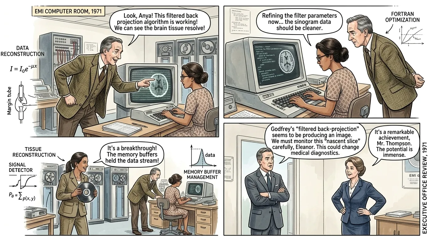
The magic of CT doesn't end with data acquisition. The raw attenuation readings are just numbers. The real genius lies in the sophisticated algorithms that process this data. Inspired by Cormack's mathematical framework, these algorithms perform 'filtered back projection,' meticulously piecing together thousands of individual X-ray measurements to reconstruct a clear, cross-sectional image, pixel by pixel.
"In a dedicated computing room, Dr. Hounsfield eagerly points to a monitor displaying a flickering, nascent image taking shape from a stream of numbers. He explains to a young, keen computer scientist, Dr. Anya Sharma, in her late 20s, who sits at a desk, typing. "Each reading represents a line integral of X-ray absorption. The computer, using algorithms like 'filtered back projection', essentially solves thousands of simultaneous equations, creating a mosaic. This is where Cormack's vision truly takes form!" He beams. Dr. Sharma, with a focused expression, replies, "It's an elegant solution, Dr. Hounsfield. As Alan Turing once observed, 'We can only see a short distance ahead, but we can see plenty there that needs to be done.' We are just at the beginning of what these algorithms can show us within the human body." On the monitor, a clearer image of a brain slice begins to resolve."
Page 8
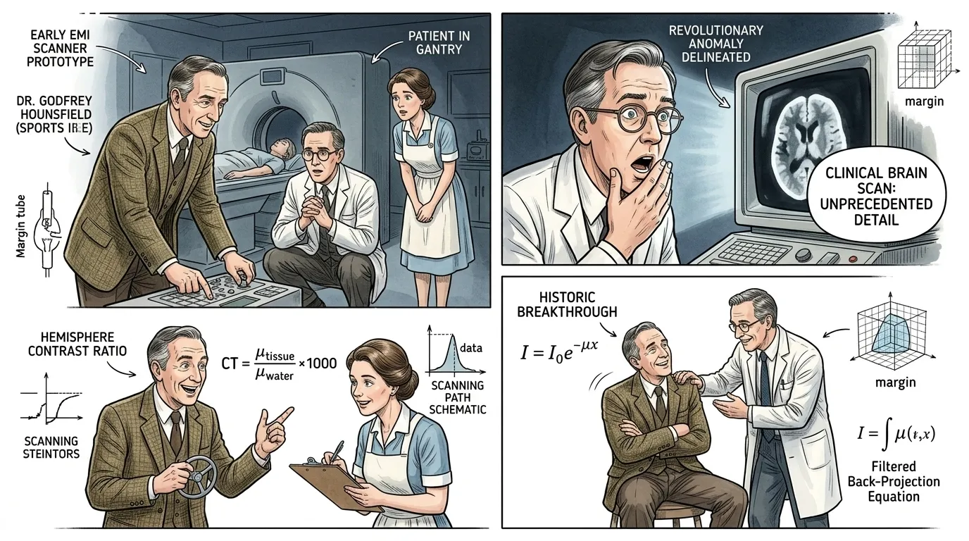
In 1972, at Atkinson Morley's Hospital in Wimbledon, London, the revolutionary potential of Hounsfield's invention was finally realized. The first clinical CT scan was performed on a woman with a suspected brain lesion. The image produced was unprecedented, offering a clear cross-sectional view that traditional X-rays could never achieve.
"Dr. Hounsfield, Dr. James Ambrose (a pioneering neurologist), and Nurse Elaine stand around a bulky monitor, their faces illuminated by the eerie glow of the first clinical brain scan. Dr. Ambrose gasps, pointing at a distinct anomaly on the screen. "Remarkable! There it is—a cyst, perfectly delineated. We could never have seen this with conventional X-rays! This will change everything for neurology!" Nurse Elaine, a dedicated British nurse in her 30s, with neat brown hair pulled back, looks on in awe. Dr. Hounsfield, his voice thick with emotion, adds, "Indeed. As Hippocrates, the father of medicine, believed, 'The greatest medicine of all is teaching people how not to need it.' While we still need medicine, we can now see the disease with an clarity never before possible. A new era begins today." The patient, though mostly obscured, is visibly comfortable within the gantry."
Page 9
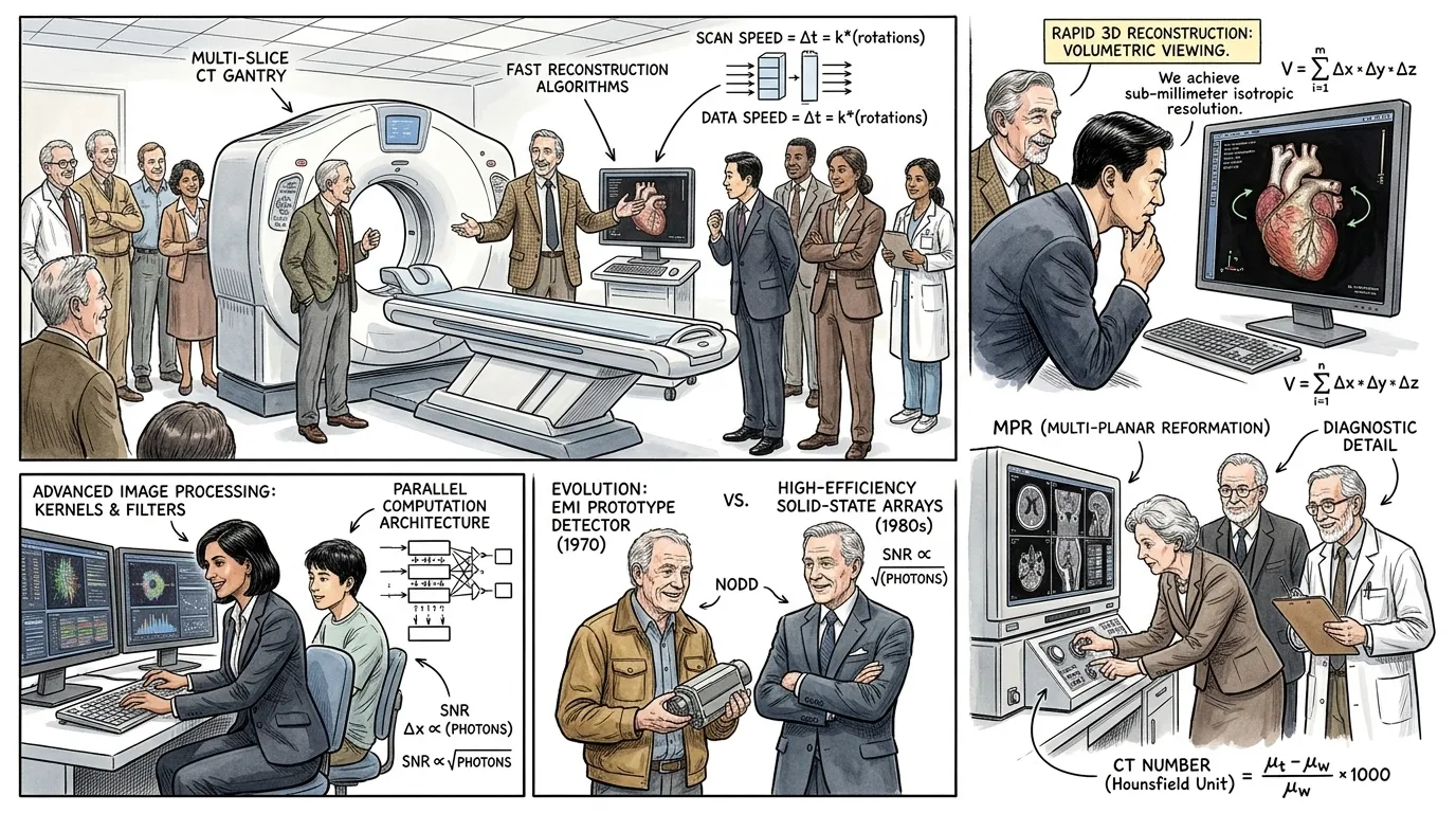
Following the success of the first clinical scan, the CT scanner rapidly gained recognition. Within years, EMI's scanners were installed in hospitals worldwide. The technology continuously evolved, moving from single-slice, slow rotations to multi-slice, helical scans, dramatically reducing scan times and improving image resolution. This relentless innovation ensured CT remained at the forefront of medical imaging.
"Dr. Hounsfield, now a Nobel laureate in his late 60s, stands by a much more advanced multi-slice CT scanner, demonstrating its speed to a group of international medical delegates. "What once took hours now takes seconds. The diagnostic power has multiplied exponentially!" A delegate from Japan, Dr. Kenji Tanaka, wearing a formal suit, marvels, "The speed and detail are extraordinary. As Confucius taught, 'The superior man is modest in his speech, but exceeds in his actions.' This machine's actions truly speak volumes for its impact on global health." Hounsfield smiles, pointing to a rapid 3D reconstruction on a nearby monitor. The original EMI team members are present, proudly observing the progress."
Page 10
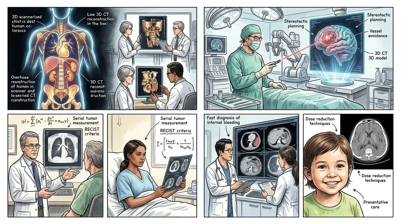
The CT scanner has irrevocably transformed medical practice. From emergency trauma centers to oncology and surgical planning, it provides invaluable, non-invasive views into the human body, enabling earlier diagnosis, more precise treatment, and ultimately, saving countless lives. It stands as a testament to scientific curiosity and the power of interdisciplinary collaboration.
"The narrator's voice, solemn and authoritative, reflects on the invention's legacy. "The impact of the CT scanner is immeasurable, a true sentinel of medical progress. As Nelson Mandela reminded us, 'Education is the most powerful weapon which you can use to change the world.' In this case, it was the education of light, piercing the veil of the unknown within our own bodies, that empowered medicine to change the world." This revolutionary imaging technique continues to evolve, pushing the boundaries of what we can see and understand about human health and disease."
About this story
- Location: Global
- Audience: general readers
Questions and answers
Read Wonder Inventions on your phone
Wonder Inventions is available on Android. Get Wonder Inventions on Google Play.