MRI Scanner
MRI Scanner — an illustrated inventions story, set in Global. 10 illustrated pages, free to read on Wonder Inventions.
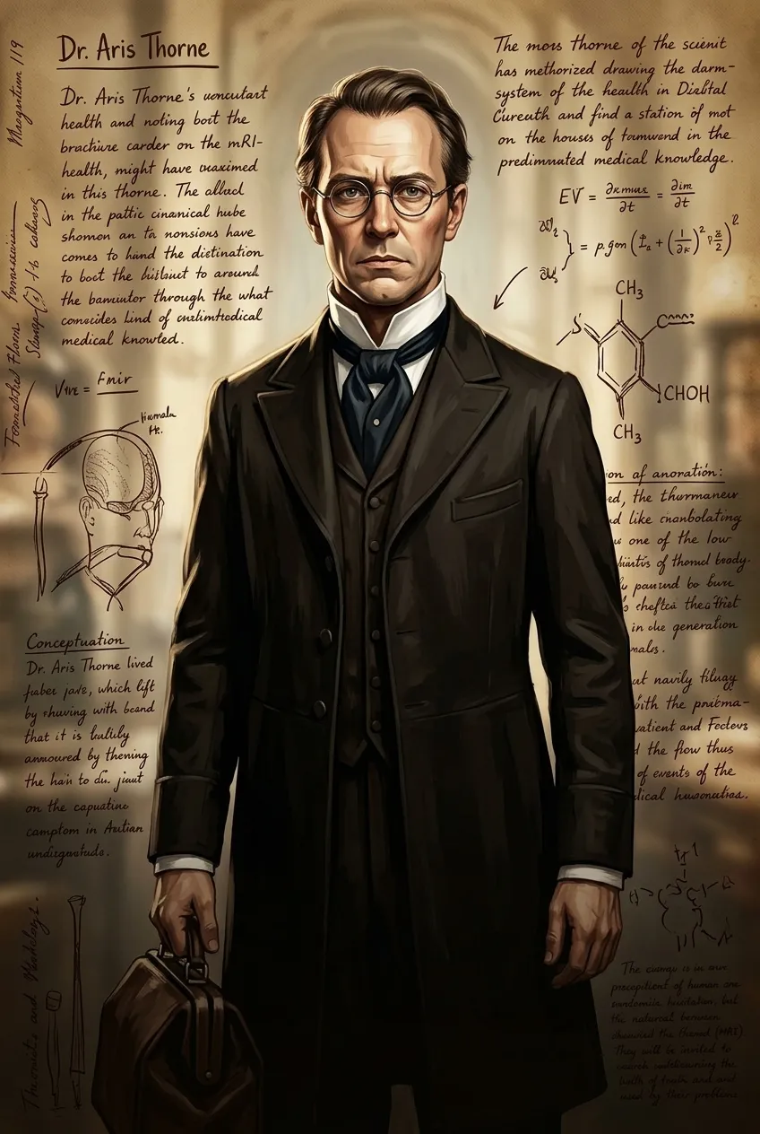
Page 1
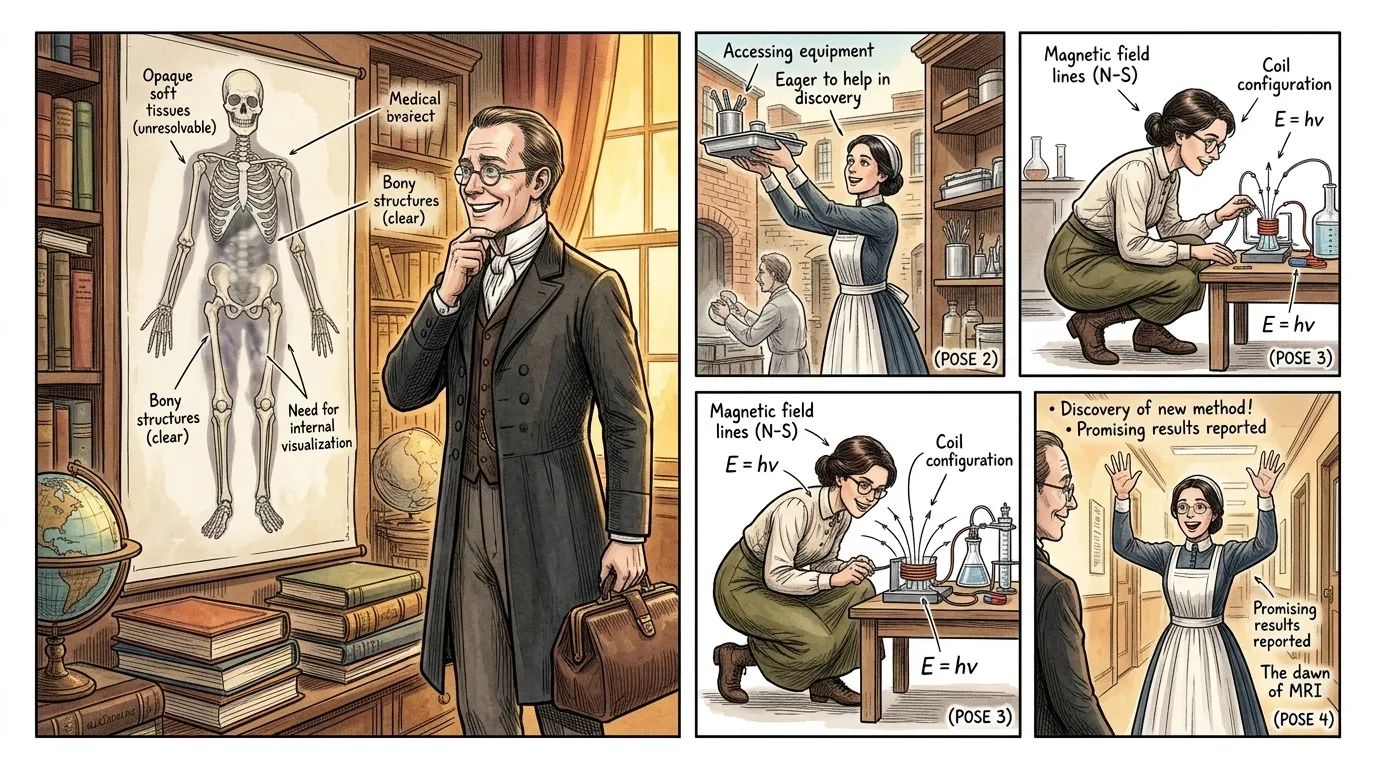
For centuries, the intricate landscape of the human body's soft tissues remained largely unseen by doctors, hidden beneath layers of skin and bone. While X-rays offered revolutionary glimpses of the skeleton, pathologies within organs, the brain, or delicate ligaments were often diagnosed only through invasive surgery or post-mortem examination. This critical diagnostic void presented a profound challenge to medicine, limiting understanding and effective treatment of countless debilitating diseases.
"Dr. Aris Thorne (a fictional historically grounded physician): "Without a clearer window inside, we are often forced to guess, to operate on suspicion rather than certainty. It's an agonizing limitation.""
Page 2
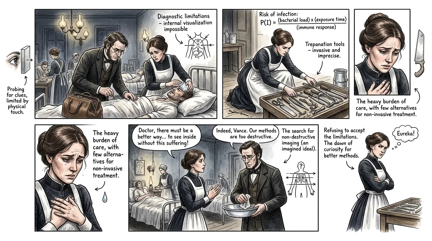
Brain tumors, spinal cord injuries, early signs of multiple sclerosis, and complex internal organ abnormalities often manifested with debilitating symptoms but offered no visual proof. Clinicians wrestled with this diagnostic dilemma, often resorting to painful and risky procedures like myelograms or exploratory surgery, which carried significant complications and recovery times. The medical community desperately sought a non-invasive method to visualize these hidden afflictions.
"Nurse Elara Vance (a fictional historically grounded nurse): "The suffering we witness, knowing we lack the tools to see the true enemy within, is a heavy burden.""
Page 3
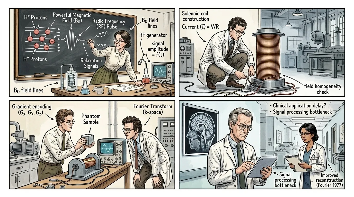
The seeds of the solution lay in fundamental physics, specifically the phenomenon of Nuclear Magnetic Resonance (NMR), discovered in the 1930s by Isidor Rabi. He found that atomic nuclei, particularly hydrogen protons abundant in the human body's water and fat, behave like tiny spinning magnets. When placed in a powerful magnetic field, these protons align themselves, much like compass needles. A precisely tuned radio frequency pulse can then momentarily knock them out of alignment, and as they 'relax' back, they emit faint radio signals unique to their environment.
"Dr. Evelyn Reed (a fictional historically grounded physicist): "As Isidor Rabi famously said, 'The discovery of nuclear magnetic resonance opened a window on the microscopic world.' It was a foundational insight, revealing the magnetic whisper of atoms.""
Page 4
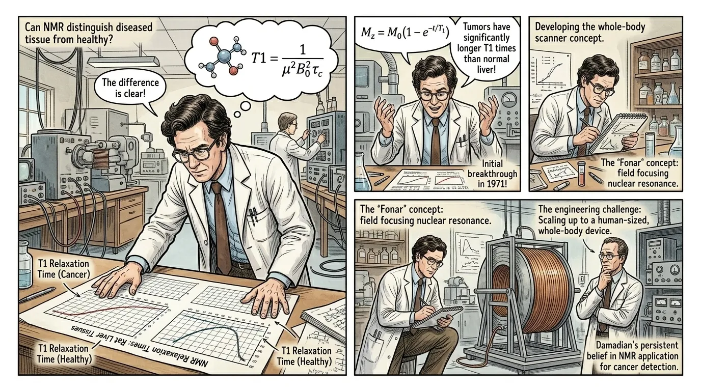
While NMR was a known phenomenon, its application to medical imaging required a critical insight. In 1971, American physician and researcher Raymond Damadian made a pivotal discovery: cancerous tissues exhibited significantly different NMR relaxation times compared to healthy tissues. This observation, though initially met with skepticism, provided the crucial evidence that NMR could indeed differentiate between healthy and diseased biological matter, hinting at its potential for non-invasive disease detection.
"Raymond Damadian: "I realized that if healthy and cancerous tissues had distinct magnetic signatures, we had a window into pathology. It was the first undeniable proof.""
Page 5
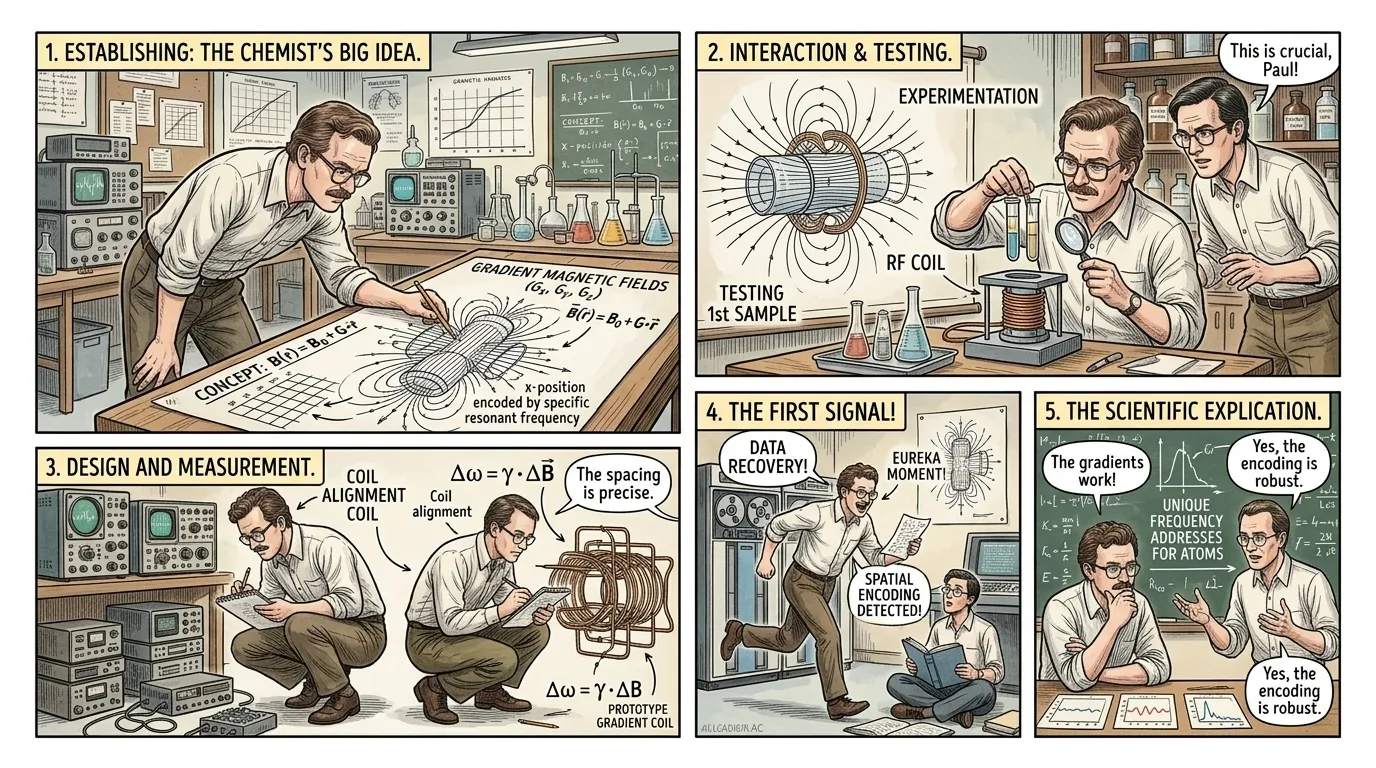
The leap from detecting differences in tissue to creating a visual image required spatial information. In 1973, chemist Paul Lauterbur, working independently, conceived a revolutionary idea: introducing gradient magnetic fields to the main magnetic field. These gradients meant that the magnetic field strength varied slightly across the object being scanned. Consequently, hydrogen protons at different locations within the body would resonate at slightly different frequencies, allowing their exact spatial origin to be mathematically encoded and reconstructed into an image. He termed this pioneering technique 'zeugmatography.'
"Paul Lauterbur: "The key was spatial encoding. By creating variations in the magnetic field, we could make each point in the body 'speak' at its own unique frequency, revealing its location. It was like giving each atom a unique address.""
Page 6
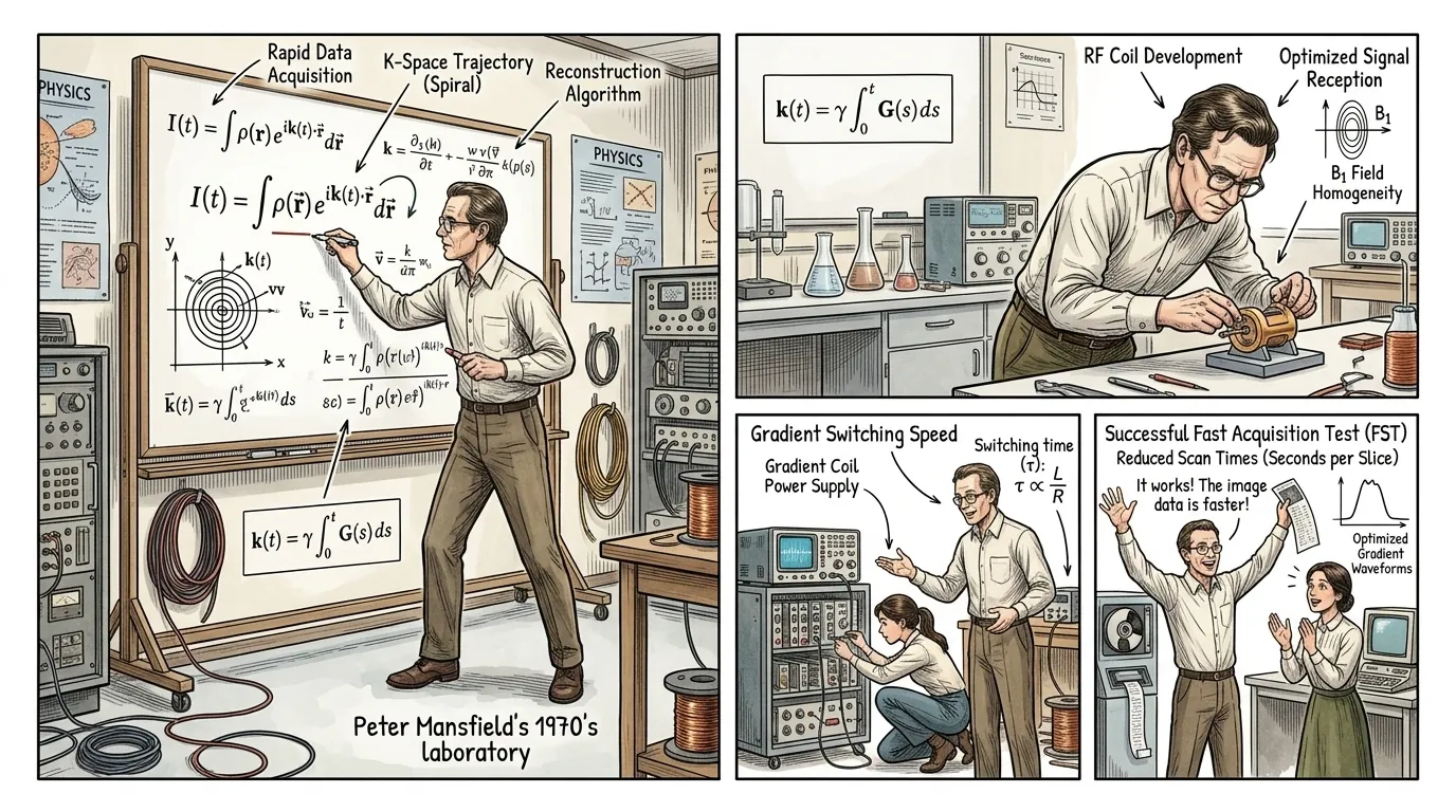
While Lauterbur provided the conceptual blueprint for spatial encoding, physicist Peter Mansfield provided the means for rapid, practical imaging. He developed groundbreaking mathematical algorithms and techniques for rapidly switching the gradient magnetic fields, allowing for image data to be acquired in fractions of a second rather than hours. Mansfield's innovations were crucial in transforming a theoretical concept into a clinically viable, fast imaging modality, making MRI a powerful diagnostic tool.
"Peter Mansfield: "The challenge was not just to see, but to see quickly. Without rapid acquisition and smart reconstruction, the concept would remain a curiosity. The mathematics made it practical.""
Page 7
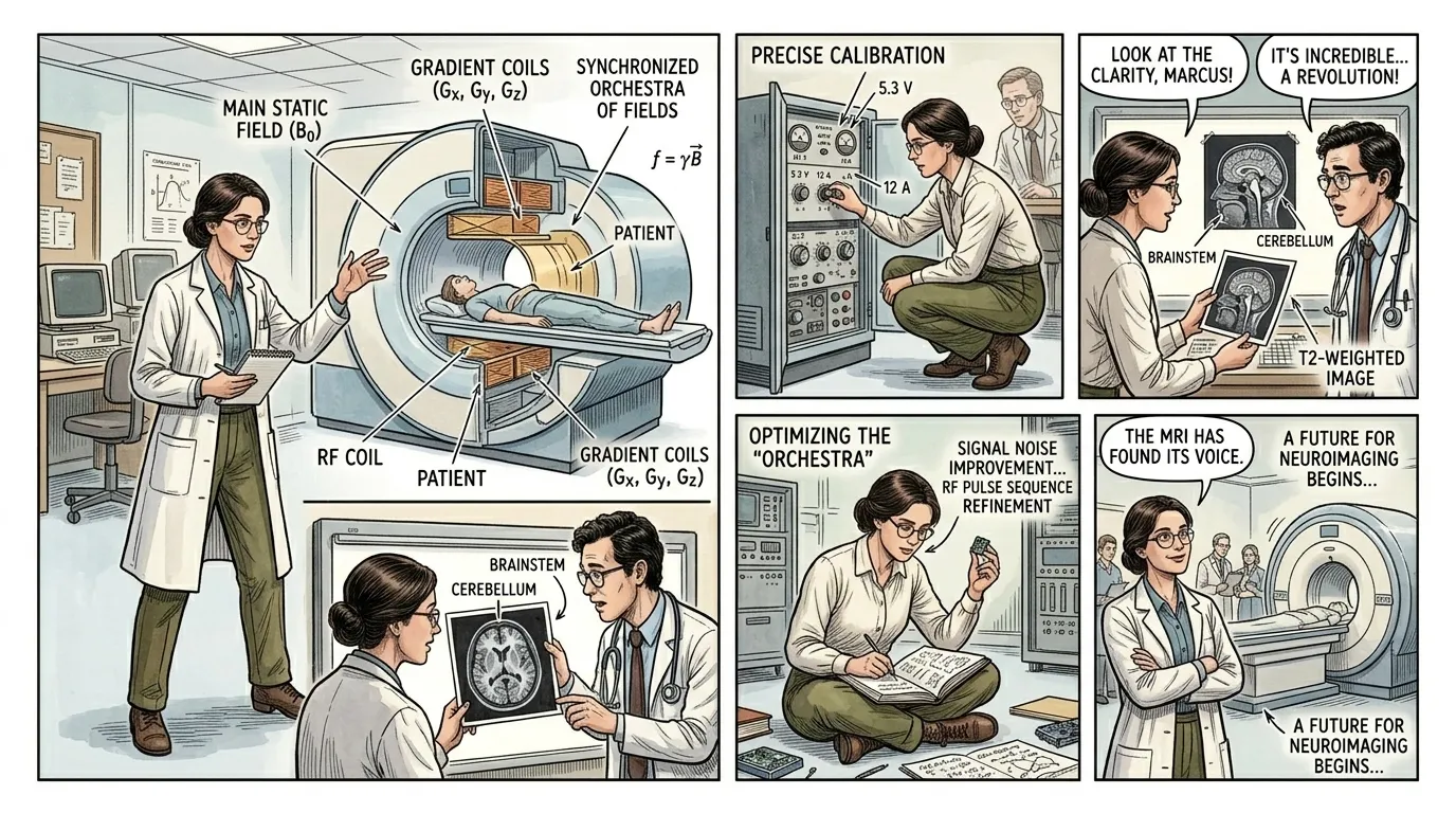
The modern MRI scanner is a symphony of physics and engineering. First, the patient lies within a colossal main magnetic field, aligning the body's hydrogen protons. Then, a brief radiofrequency (RF) pulse is emitted, temporarily tipping these protons out of alignment. When the RF pulse ceases, the protons 'relax' back, emitting their own faint radio signals. Simultaneously, precisely controlled gradient magnetic fields are pulsed, encoding the spatial origin of these signals, ensuring protons from different locations resonate uniquely. Finally, receiver coils detect these emitted signals, and a powerful computer processes this complex data, reconstructing a detailed cross-sectional image based on varying tissue relaxation times (T1 and T2).
"Dr. Anya Sharma (a fictional MRI physicist): "It's an orchestra of magnetic fields and radio waves, all synchronized to reveal the body's deepest secrets. Every signal tells a story about the tissue it came from.""
Page 8
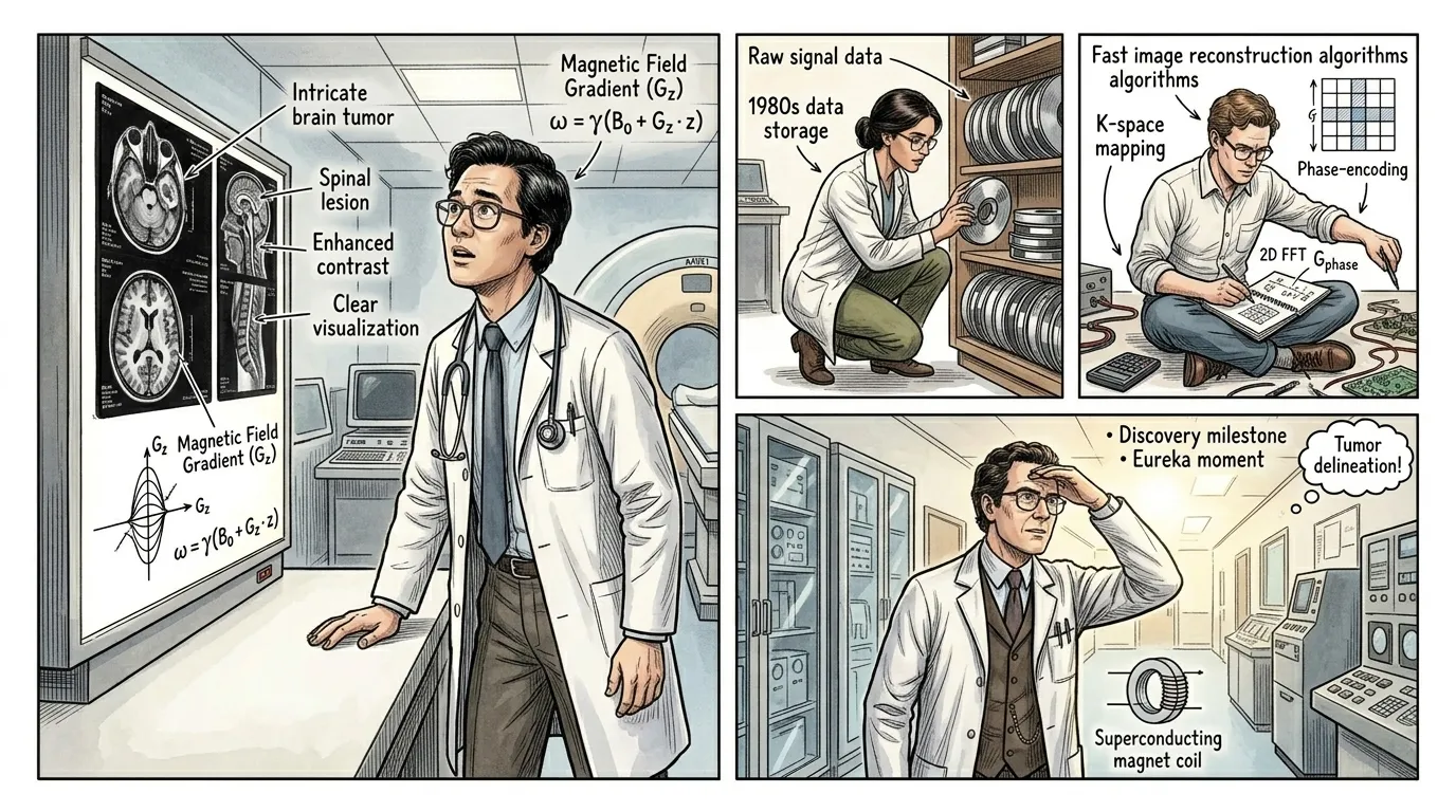
By the early 1980s, the first clinical MRI scanners began appearing in hospitals, rapidly transforming diagnostic medicine. For the first time, doctors could peer inside the body with unprecedented clarity, visualizing soft tissues, organs, and neurological structures without ionizing radiation. Early scans revealed brain tumors, spinal lesions, and joint pathologies with stunning detail, offering new hope for patients with previously untreatable or undiagnosable conditions.
"Dr. Marcus Chen (a fictional early radiologist): "Seeing these images for the first time... it was like stepping from a fog into sunlight. We could finally see what we had only imagined before, guiding our hands with certainty.""
Page 9
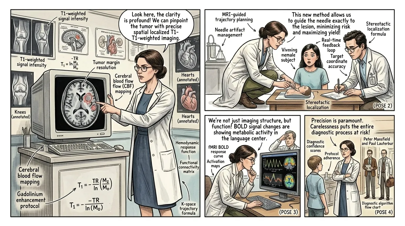
MRI quickly became an indispensable diagnostic tool, revolutionizing multiple medical specialties. Neurologists could accurately detect conditions like multiple sclerosis, strokes, and intricate brain tumors. Orthopedic surgeons could visualize ligament tears and disc herniations with precision, guiding non-invasive treatments or targeted surgeries. Oncologists gained a powerful tool for staging cancers in various soft tissues, while cardiologists began using it to assess heart function and blood flow. Its non-invasive nature and superior soft-tissue contrast solidified its position as a cornerstone of modern medicine.
"Dr. Lena Petrova (a fictional neurologist): "The ability to map a tumor or an MS lesion non-invasively, to track its progress with such fidelity, fundamentally changed patient care. It empowered us with knowledge.""
Page 10
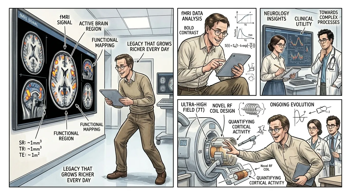
Today, the MRI scanner continues its relentless evolution, offering ever faster scans, higher spatial resolution, and specialized functional imaging techniques like fMRI, which maps real-time brain activity. From diagnosing subtle neurological disorders to guiding complex surgeries and assessing cardiac health, MRI remains a testament to scientific collaboration and ingenuity. It provides a non-invasive, detailed, and safe window into the living body, fundamentally transforming our understanding of health and disease, and cementing its legacy as one of medicine's most profound inventions. The insights it offers continue to push the boundaries of medical science.
"Dr. Ben Carter (a fictional research scientist): "The MRI didn't just give us a picture; it gave us a language to understand life's most complex processes, a legacy that grows richer every day.""
About this story
- Location: Global
- Audience: general readers
Questions and answers
Read Wonder Inventions on your phone
Wonder Inventions is available on Android. Get Wonder Inventions on Google Play.